Grant Smith, DDS
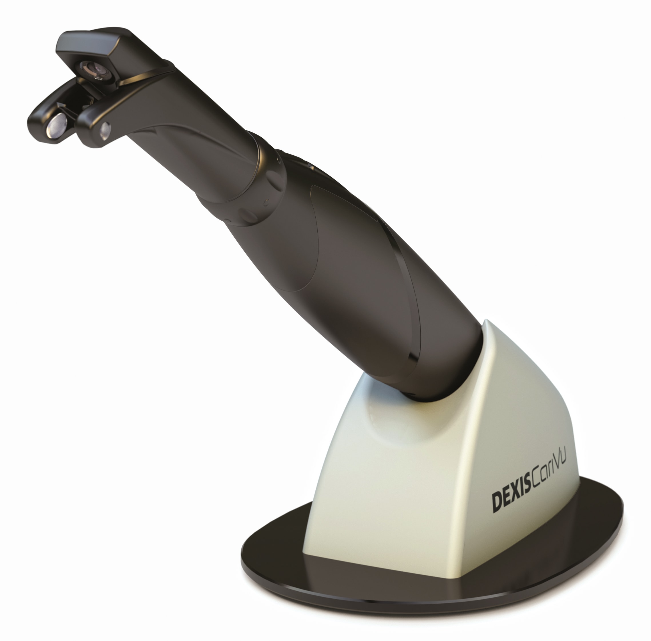
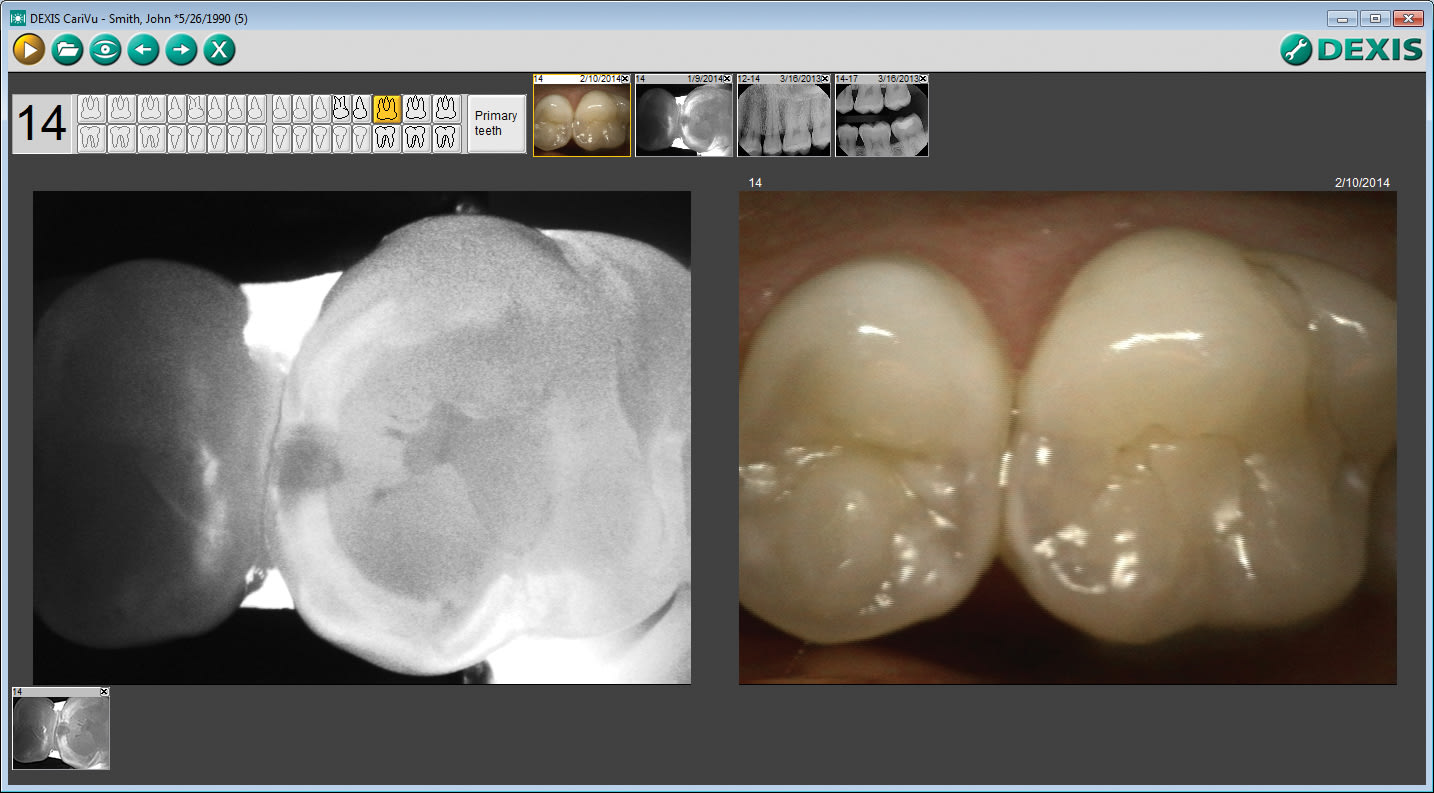
While many new technologies interest me, the CariVu caries detection device (Figure 1) has become a perfect addition to my imaging tools. This device uses near-infrared (NIR) light that illuminates both sides of the tooth simultaneously to detect carious lesions and cracks interproximally, occlusally, and around cracks and restorations. CariVu’s technology is different than other types of caries detection devices—some show results through a range of colors or a number that correlates to a degree of mineralization. On a CariVu image (Figure 2), healthy parts of the tooth appear light and lesions appear dark because healthy structures reflect the NIR light, whereas the porous carious lesions trap and absorb the light.
What Makes this Device Different
The CariVu device yields an understandable image without the need to clean the tooth of bacteria, calibrate the device, or become versed in the meaning of multiple color codes or numeric indicators. The NIR technology eliminates these extra steps, making caries detection that much faster and simpler for the clinician and the patient.
This technology has helped me with diagnosis and patient education, and is easy to use. A rubberized tip hugs the tooth to illuminate it from the buccal and lingual aspects for the capture of a clear picture. I can easily move the tip around, pivot it for an active visualization of the field, and change the focal depth as I move across the tooth to get a better 3-dimensional concept of the tooth. After I transilluminate the patient’s tooth, I can capture the image and enlarge it as necessary to show the patient areas of concern.
Even patients who are not receptive to low-dose digital x-rays can benefit because CariVu technology does not involve radiation. While it does not eliminate the need for x-rays, CariVu is a helpful addition to other imaging technologies, in some cases changing my expectations. With this device, I know the exact location and extent of the decay before opening the tooth. Sometimes for various reasons, x-rays have been misleading. I have opened teeth that I thought might have significant caries around a fracture and found little to none, and I have found teeth where I expected little decay around a fracture that had substantial caries.
According to a research study, CariVu technology has an interproximal dentin caries detection rate of 99%.1 This accuracy gives me a sense of security because I know the size and scope of decay and the extent of fractures that I would not have otherwise seen on an x-ray. Therefore, I am better prepared to excavate and restore the space. Being able to see the correlated size, shape, and scope of initial carious lesions, as well as around margins of failing restorations, lets me effectively plan treatment and allows for smaller accesses and less invasive treatment.
About the Author
Grant Smith, DDS, attended dental school at the University of Missouri-Kansas City (UMKC), and completed a residency in the special patient care clinic. He worked with transplant patients, oncology patients, and others who needed more care than the rank and file at UMKC could provide, or they needed things done quickly to prepare for surgery. Dr. Grant learned more in this residency than he has perhaps ever learned in his whole life. He is the “Gentle Dentist” at Prairie Village Dentists in Prairie Village, Kansas.
Reasons to Buy CariVu Caries Detection Device
Safe and Advantageous
Does not emit ionizing radiation so even patients who are not receptive to low-dose digital x-rays can benefit from it
Accurate
99% accuracy rate for interproximal caries
Small and Portable
Compact, portable, and easy to use
Comprehensive
Together with other imaging technologies, offers a comprehensive image history of the health of a patient’s teeth
DEXIS, LLC
888-883-3947
www.dexis.com
References
1. Kühnisch J. Study project “benefits of the DIAGNOcam procedure for the detection and diagnosis of caries:” summary of the final report. Kavo Kerr Group website. www.diagnocam.com/img_cpm/350_DIAGNOcam/files/DIAGNOcam_LMUAbschlussbericht-en.pdf. Accessed September 11, 2015.


